BRAIN MRI and Autism

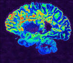
Autism and Brain MRI

Wessam Bou-Assaly, MD

A national research network led by UNC School of Medicine’s Joseph Piven, MD, found
that many toddlers diagnosed with autism at two years of age had a substantially greater
amount of extra-axial cerebrospinal fluid (CSF) at six and 12 months of age, before
diagnosis is possible. They also found that the more CSF at six months — as measured
through MRIs — the more severe the autism symptoms were at two years of age.
Until the last decade, the scientific and medical communities viewed CSF as merely a
protective layer of fluid between the brain and skull, not necessarily important for
proper brain development and behavioral health. But scientists then discovered that
CSF acted as a crucial filtration system for byproducts of brain metabolism.
Every day, brain cells communicate with each other. These communications cause brain
cells to continuously secrete byproducts, such as inflammatory proteins that must be
filtered out several times a day. The CSF handles this, and then it is replenished with
fresh CSF four times a day in babies and adults.
The researchers found that increased CSF predicted with nearly 70 percent accuracy
which babies would later be diagnosed with autism. It is not a perfect predictor of autism, but the CSF differences are observable on a standard MRI. “

Meditation and Brain Structure

Effect of Meditation on the Brain

Wessam Bou-Assaly, MD


The effect of the meditation on the brain has been a subject of many researches recently. Meditation appears to have an amazing neurological benefits, suspected from ancient times, just now being confirmed by MRI, functional MRI and EEG.

Most recently, a study conducted by a Harvard affiliated team out of Massachusetts General Hospital (MGH), concluded that meditation has tangible effects, confirmed by MRI, on the human brain structure. An 8 week program of mindfulness meditation studied by MRI scans, showed  for the first time clear evidence that meditation produces “massive changes” in brain Gray Matter.

MRI scans documented for the first time how meditation produces massive changes inside the brain’s gray matter. The structural differences between the brains of experienced meditation practitioners and individuals with no history of meditation, showed thickening of the cerebral cortex in areas associated with attention and emotional integration.

Meditation also appears to help preserve the aging brain. A study from UCLA found that long-term meditators had better-preserved brains than non-meditators as they aged. Participants who’d been meditating for an average of 20 years had more grey matter volume throughout the brain.

A Yale University interesting studies in the last few years, found that mindfulness meditation also decreases activity in the default mode network (DMN), the brain network responsible for mind-wandering and self-referential thoughts – a.k.a., “monkey mind.” The DMN is active when we’re not thinking about anything in particular, wandering from thought to thought. Since mind-wandering is typically associated with being less happy, ruminating, and worrying about the past and future, it’s the goal for many people to dial it down. Several studies have shown that meditation, though its quieting effect on the DMN, appears to do just this.

A Johns Hopkins study looked at the relationship between Mindfulness Meditation and its effect on depression, anxiety, and pain. Researcher Madhav Goyal and his team found that the effect size of meditation was moderate, at 0.3 but equal to the effect size for antidepressants which is also 0.3, which makes the effect of Meditation sound pretty good, making the Meditation an active form of brain training.

Meditation has central effects on improving attention and concentration, reduces anxiety, especially social anxiety, can be very effective in helping people recover from various types of addiction, including quitting smoking and can help kids in school performance.

Wessam Bou-Assaly, MD is a Radiologist in Ann Arbor, Michigan.

Wessam Bou-Assaly, MD : The Risk of X-Ray

safety-sign-radiationThe Risk of X-rays

X-rays are produced when charged particles (electrons or ions) of sufficient energy hit a material and are suddenly decelerated upon their collision.

X-ray images are produced when a patient is placed in front of an X-ray detector and is then illuminated by short X-ray pulses. X-rays are absorbed by dense material with high atomic number such as bones, which are rich with calcium, and appear white on the resulting image. In the other hand, material with low or no atomic number, such as air in lungs, show up as dark patches on X-ray images because of their low absorption rates.

Type of X-Ray:

There are mainly 3 types of clinically used X-ray:

Radiography is the most familiar type of X-ray imaging. It is used to image mainly bones and the chest. Radiography also uses the smallest amounts of radiation.

Fluoroscopy is continuous live use of X-ray, and cane considered as a movie equivalent. The radiologist can watch the X-ray of the patient moving in real-time to watch the activity of the gut after a barium meal or intravascular contrast. Fluoroscopy uses more X-ray radiation than a standard X-ray, but the amounts are still tiny.

During Computed tomography (CT), the patient lies on a table and enters a ring-shaped scanner. A fan-shaped beam of X-rays passes through the patient into detectors placed across the patient’s body. The patient moves slowly into the machine so that a series of slices can be taken. This procedure uses the highest dose of X-rays because so many images are taken in one sitting.

The risk of X-Ray:

X-rays can cause mutations in the patient’s DNA and, therefore, might lead to cancer later in life. For this reason, X-rays are classified as a carcinogen by both the World Health Organization (WHO) and the US government. However, the benefits of X-ray technology far outweigh the potential negative consequences of using them. It is estimated that 0.4% of cancers in America are caused by CT scans and this level is expected to rise in parallel with the increased usage of CT scans in medical procedures.

Each procedure has a different risk associated with it, depending on the type of X-ray and the part of the body being imaged. In example:

  • Chest X-ray is equivalent to 2.4 days of natural background radiation
  • Lumbar spine is equivalent to 182 days of natural background radiation
  • Upper gastrointestinal barium exam is equivalent to 2 years of natural background radiation
  • CT head is equivalent to 243 days of natural background radiation
  • CT abdomen is equivalent to 2.7 years of natural background radiation.

Even though the X-ray has been associated with risk of cancer, their benefit if used properly surpass their claimed danger: the importance of making the right diagnosis and choosing the correct course of treatment makes X-rays far more beneficial than they are dangerous.

Whether there is a small risk or no risk at all, the X-rays are here to stay.

Wessam Bou-Assaly, MD is a highly experienced radiologist in Ann Arbor -Michigan with long year of practice.

Wessam Bou-Assaly – What it Takes to Succeed as a Medical Student

Medical StudentMedical school requires several years and a lot of work. Wessam Bou-Assaly graduated from medical school in 2000. He studied at a university in France and then entered a radiology residency program at Caen and Lille in France. He worked hard to become a successful radiologist and to specialize in neuroradiology and nuclear medicine.

Medical students spend four years studying in order to graduate with their degree. The first two years of medical school involve classroom study. Students learn about the fundamentals of medicine. They also study a variety of medical fields in order to develop a well-rounded knowledge of the field. The second two years involve clinical practice. They learn how to treat patients and they develop experience in working with patients.

In order to succeed as a medical student, you should work hard on your studies. It is important to focus on your studies and to develop a strong study group. It is also important to make strong connections with your medical professors. These connections may help you find a great residency after you graduate.

Successful medical students begin networking early on. This process can help students find great residency programs, fellowships, and jobs after graduating from medical school. It can be difficult to earn a spot in a medical school, and graduating requires a lot of work and a lot of time. Wessam Bou-Assaly is a radiologist who worked hard to graduate from medical school. After earning his degree, he completed a radiology residency and fellowship programs in neuroradiology and nuclear medicine.


Wessam Bou-Assaly – Nuclear Medicine versus Radiology

diagnostic_radiology_slide1
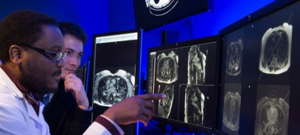
Nuclear medicine is a sub discipline of radiology. Wessam Bou-Assaly is a radiologist who specializes in nuclear medicine. In 2007, he completed a fellowship program in nuclear medicine. He is a member of the Society of Nuclear Medicine, and he has conducted a large amount of research in the field. Nuclear medicine is different from radiology.

Radiology involves using X-ray imaging to diagnose and treat diseases, injuries, and illnesses. Radiologists use a wide array of imaging techniques including computed tomography (CT), ultrasound, MRI and X-ray radiography. These professionals create images by projecting X-rays over the body. Machines measure where the rays pass through, and where they are stored in the body. That information is then used to create an image of the human body.

Nuclear medicine is a type of radiology, however, it uses a very different method to create images. Professionals introduce small amounts of radioactive substances into their patients’ bodies. These substances are either injected or ingested. The radiologist will then use gamma cameras to form images based on the radiation that is emitted from the body.

Nuclear medicine is different from other types of radiological images, because the images show physiological functions. Radiologists can use nuclear medicine to study the flow of blood to the brain or the function of the kidneys. Other forms of radiological imaging, such as CT scans or MRI scans, only create an image.

Wessam Bou-Assaly is a radiologist who has studied neuroradiology and nuclear medicine. He is knowledgeable and experienced in his field.

Sources: http://interactive.snm.org/docs/whatisnucmed2.pdf