Autism and Brain MRI
Wessam Bou-Assaly, MD
A national research network led by UNC School of Medicine’s Joseph Piven, MD, found
that many toddlers diagnosed with autism at two years of age had a substantially greater
amount of extra-axial cerebrospinal fluid (CSF) at six and 12 months of age, before
diagnosis is possible. They also found that the more CSF at six months — as measured
through MRIs — the more severe the autism symptoms were at two years of age.
Until the last decade, the scientific and medical communities viewed CSF as merely a
protective layer of fluid between the brain and skull, not necessarily important for
proper brain development and behavioral health. But scientists then discovered that
CSF acted as a crucial filtration system for byproducts of brain metabolism.
Every day, brain cells communicate with each other. These communications cause brain
cells to continuously secrete byproducts, such as inflammatory proteins that must be
filtered out several times a day. The CSF handles this, and then it is replenished with
fresh CSF four times a day in babies and adults.
The researchers found that increased CSF predicted with nearly 70 percent accuracy
which babies would later be diagnosed with autism. It is not a perfect predictor of autism, but the CSF differences are observable on a standard MRI. “

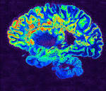
 Brain imaging with MRI, SPECT, and PET can improve diagnostic accuracy in differentiating Alzheimer Disease (AD) from potentially treatable causes of dementia such as toxic metabolic states, depression, and normal pressure hydrocephalus. When PET results are combined with clinical criteria, the false positive rate in AD can be reduced from 23% to 11%.[9] The classic pattern of AD on PET imaging is bilateral temporoparietal and posterior cingulate cortex hypometabolism; abnormal metabolism can also be seen asymmetrically, particularl yearly in the disease. Frontal lobe involvement may also be seen in later stages. The exact cause for the decline in brain glucose metabolism in AD remains unclear. Hippocampal atrophy may be seen on conventional cross-sectional imaging.